
ISSN 0001-6012 eISSN 2215-5856
Acta Médica Costarricense / 2026 / enero-marzo; 68 (1): 1-7
doi: 10.51481/amc.v68i1.1525

Clinical Management of a Mesoamerican Rattlesnake (Crotalus simus) Envenomation in Costa Rica
(Manejo clínico de un accidente ofídico por serpiente de cascabel mesoamericana (Crotalus simus) en Costa Rica)
Felipe Triana1, Stanley Grant Villegas2, José L. Ulloa Méndez3, Fabián Bonilla4
Afiliación Institucional:
1Universidad de Costa Rica, Facultad de Microbiología, Instituto Clodomiro Picado, San José, Costa Rica; Universidad de Costa Rica, Sistema de Estudios de Posgrado, Programa de Posgrado en Biología, San José, Costa Rica; University of Wollongong, School of Science, Environmental Futures Research Center, Wollongong, Australia.
 0000-0003-2416-3616
2Caja Costarricense de Seguro Social, Hospital Dr. Rafael Ángel Calderón Guardia, Departamento de Medicina, Servicio de Medicina Interna, San José, Costa Rica.
0009-0006-6373-3975
3Reserva Conchal, Trabajador Independiente, Santa Cruz, Guanacaste, Costa Rica.
0009-0002-2827-4399
4Universidad de Costa Rica, Facultad de Microbiología, Instituto Clodomiro Picado, San José, Costa Rica.
 0000-0002-5095-2750
Abreviaturas:
C. simus ; Crotalus simus.
h; Horas.
ICP; Instituto Clodomiro Picado.
CCSS; Caja Costarricense de Seguro Social.
VAC; Vacuum-assisted closure therapy.
Fuentes de apoyo: No hay.
Declaración de interés: Los autores declaran que no existe ningún conflicto de intereses que pueda percibirse como perjudicial para la imparcialidad de este manuscrito.
Abstract
Snakebite envenomation is a relevant public health problem in Costa Rica, a country with high snake diversity and an annual incidence of 12 to 19 cases per 100,000 inhabitants. However, due to the national production of antivenom, its universal availability in public hospitals, and decades of research on snake envenomation, morbidity and mortality rates are low. Despite representing approximately 1% of cases in the country, envenomations caused by Crotalus simus are clinically relevant due to their severity, the broad geographic distribution of this species, and abundance of specimens. We present a case report of a snakebite envenomation caused by an adult C. simus (Mesoamerican rattlesnake) in northwestern Costa Rica. The patient was a 40-year-old man weighing 100 kg who was bitten on the right hand with both fangs. Following the event, the patient was transferred to Hospital de la Anexión de Nicoya, where he received 20 vials of PoliVal-ICP antivenom. The main clinical manifestations included local pain, edema, erythema, and paresthesia. In addition, laboratory tests revealed hematological and coagulation abnormalities consistent with the venom composition of adult individuals of this species, which is rich in snake venom metalloproteinases. On the second day, the patient was transferred to Hospital Calderón Guardia, a higher-level medical facility in the capital city of San José, where he was reevaluated due to persistent edema. On the third day, the medical team decided to perform a fasciotomy and initiate antibiotic therapy. In the following days, the patient underwent surgical washouts and vacuum-assisted closure therapy. Additionally, the patient developed serum sickness on the sixth day. On the tenth day, the VAC system was removed, and the patient remained under observation until day 14, when he was discharged. Finally, the patient attended subsequent rehabilitation therapy sessions to restore motor function. This case highlights the importance of timely hospital transfer and early antivenom administration as key strategies to reduce morbidity and mortality associated with snakebite envenomation. However, it also demonstrates the need to strengthen surveillance and standardize national hospital protocols to prevent unnecessary surgical interventions.
Descriptores: antivenom, fasciotomy, snake bite, Crotalus, rattlesnake venom, snake venom
Resumen
El accidente ofídico es un problema de salud pública relevante en Costa Rica, país que cuenta con una alta diversidad de serpientes y una incidencia anual de 12 a 19 casos/100,000 habitantes. Sin embargo, gracias a la producción nacional de suero antiofídico, su disponibilidad universal en hospitales públicos y décadas de investigación en envenenamientos por estos animales, las tasas de morbilidad y mortalidad son bajas. A pesar de representar aproximadamente el 1% de los casos en el país, los envenenamientos por Crotalus simus son clínicamente relevantes debido a su gravedad, la amplia distribución geográfica de la especie y la abundancia de especímenes. Presentamos un reporte de caso de un accidente ofídico causado por un adulto de C. simus (serpiente de cascabel mesoamericana) en el noroeste de Costa Rica. El paciente fue un hombre de 40 años, con un peso de 100 kg, quien fue mordido con ambos colmillos en la mano derecha. Posterior al evento, el paciente fue trasladado al Hospital de la Anexión de Nicoya, donde se le administraron 20 viales de suero antiofídico PoliVal-ICP. Entre los síntomas principales se reportaron dolor local, edema, eritema y parestesia. Adicionalmente, los exámenes de laboratorio revelaron alteraciones hematológicas y de la coagulación, consistentes con la composición del veneno de los adultos de esta especie, el cual es rico en metaloproteinasas. En el segundo día, el paciente fue trasladado al Hospital Calderón Guardia, un centro médico terciario con mayor capacidad resolutiva en la ciudad capital de San José, donde fue reevaluado debido a la persistencia del edema. Al tercer día, el equipo médico decidió realizar una fasciotomía e iniciar terapia antibiótica. En los días posteriores, el paciente fue sometido a lavados quirúrgicos y a la implementación de terapia de cierre asistido por vacío. Además, el paciente presentó enfermedad del suero al sexto día. El décimo día se retiró el sistema asistido por vacío y el paciente permaneció en observación hasta el día 14, cuando fue dado de alta. Finalmente, el paciente asistió luego a sesiones de terapia de rehabilitación para la recuperación de la funcionalidad motora. Este caso resalta la importancia del traslado hospitalario oportuno y la administración temprana del antiveneno como estrategias clave para reducir la mortalidad y la morbilidad asociadas al accidente ofídico. Sin embargo, también demuestra que es necesario fortalecer la vigilancia y la estandarización de los protocolos hospitalarios nacionales para prevenir intervenciones quirúrgicas innecesarias.
Keywords: antiveneno, fasciotomía, mordedura de serpiente, Crotalus, veneno de cascabel, veneno de serpiente
Fecha de recibido: 16, enero, 2026 Fecha de aceptado: 09, julio, 2026
Snakebite envenomation is a relevant disease that affects between 1.8 and 2.7 million people worldwide, causing approximately 100.000 deaths and about 400.000 physical or psychological sequelae per year.1-3 Costa Rica reports approximately 500 cases of snakebites annually, representing 12 to 19 cases/100,000 inhabitants.4,5 The high incidence observed in this small country could be explained by a great snake diversity,6 which includes 147 species, 25 of which are medically important. Despite its high incidence, the mortality rate has drastically decreased in recent decades, reaching less than 0.2 deaths/100,000 inhabitants per year.7,8 The main factors contributing to the low mortality rate from snakebite envenomation in Costa Rica are the national production of antivenom by the Instituto Clodomiro Picado (ICP) at the University of Costa Rica (https://www.icp.ucr.ac.cr/), which ensures immediate availability across the country, and the geographic accessibility together with the universal health coverage provided by the public healthcare system through the Costa Rican Social Security Fund (Caja Costarricense de Seguro Social (CCSS), https://www.ccss.sa.cr/).4,6
Although Bothrops asper causes the vast majority of snakebite envenomation in Costa Rica and is considered the most dangerous snake in the region,6,7 Crotalus simus (C. simus), is also one of the species of greatest medical relevance in the country due to (1) the severity of its envenomations despite its low incidence,9 and (2) its distribution that includes places where B. asper is not found.10
C. simus, commonly known as the Mesoamerican rattlesnake, is a large tropical viper distributed from southern Mexico to northern Costa Rica, accounting for approximately 1% of snakebite cases in the country.10 It primarily inhabits seasonal dry tropical forests, feeds on small mammals and lizards, and reproduces seasonally, where births are synchronized with the onset of the rainy season.
Studies in Costa Rica have offered initial characterizations of C. simus envenomation10 and the species’ venom composition is well studied.11,12 However, clinical reports for this species are lacking, despite it being one of the most medically relevant snakes where it occurs.9 Moreover, individuals of the Mesoamerican rattlesnake are common throughout its distribution, highlighting the importance of generating information for the region on the clinical manifestations of envenomation by this species. Therefore, we report here the case of a man who survived a severe envenomation by an adult C. simus in Costa Rica, with emphasis on his in-hospital management.
Case report
In Nosara Beach, Guanacaste, northwestern Costa Rica, on May 18, 2024, at 16:30 hours (h), a 40-year-old male patient (100 kg) was bitten by a male C. simus measuring 140.5 cm in total length (125 cm snout–vent length + 15.5 cm tail) and weighing 1650 g, while handling the snake (Figure 1). The medical history of the patient includes dyslipidemia, arterial hypertension and glaucoma, and his treatment was gemfibrozil 600 mg BID, enalapril 20 mg daily, amlodipine 5 mg daily, timolol and latanoprost ophthalmic when the bite occurred. The bite was inflicted with both fangs on the man’s right hand between the proximal phalange of the index finger and the second metacarpal bone (Figure 1). Envenomation was confirmed by the presence of two puncture marks at the fang entry site and severe local pain that quickly spread throughout the rest of the arm. Immediately after the bite, the patient was transported by ambulance to Hospital de la Anexión de Nicoya, about an hour and a half from Nosara. Within the first 30 minutes, the patient developed diaphoresis, and by approximately one hour, he exhibited sphincter relaxation. The patient lost consciousness prior to hospital admission, although he was respiring normally and did not require intubation nor an oxygen mask.
The patient arrived at the emergency department of the local hospital at 17:45 h, where physician reported hypotension (92/51 mmHg), mild hypothermia (35.4 °C) and a normal heart rate (64 bpm), O2 saturation 93%, and respiratory rate 18 bpm. Local manifestations include pain, edema, erythema and paresthesia. Initial blood tests suggested hemoconcentration, enlarged erythrocytes with increased hemoglobin content, enlarged platelets, leukocytosis and abnormalities in coagulation tests (Table 1). Fibrinogen testing was not performed. Following the protocols for snakebites established in Costa Rica by ICP and CCSS (https://www.icp.ucr.ac.cr/en/information-and-materials/information-healthcare-professionals), an initial dose of 10 vials (10 mL) of the antivenom specific against Central and South American vipers PoliVal-ICP (https://www.icp.ucr.ac.cr/en/productos/polival-icp) was administered at 1.5 h post-bite (18:00 h). Around 6 hours after the bite (22:16 h), a second dose of 10 vials was infused, which may have prevented the swelling from worsening and reduced the risk of tissue damage.
On the second day, laboratory tests showed modestly elevated creatine phosphokinase (CPK) levels (Table 1), suggesting muscle tissue injuries. Edema and bullae were present along the right upper limb (Figure 2). The doctors made the decision to transfer the patient to Hospital Calderón Guardia, a higher-level medical facility in the capital city of San José. On admission to the hospital, orthopedic surgeons evaluated the patient indicating soft tissue damage in the right forearm up to the elbow without evidence of bone fracture or other bone damage. At 21:30 h the reconstructive surgery service also evaluated the patient and found no signs of compartment syndrome. The arm was raised to reduce inflammation, and a splint was placed with wrist extension.
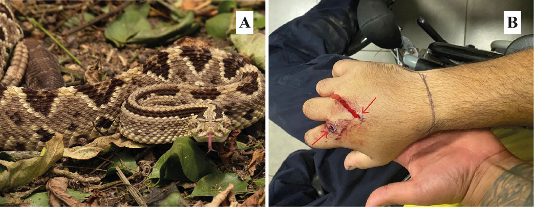
Figure 1. A) Example of a Mesoamerican rattlesnake (Crotalus simus) from the same locality as the individual involved in the incident (Nosara, Guanacaste, Costa Rica). B) Bite wound with two fang punctures on the dorsum of the right hand. Red arrows point to the fang punctures.
___
|
Table 1. Laboratory test results obtained during the hospitalization of a 40-year-old male patient following a snakebite by an adult Crotalus simus in Nosara, Guanacaste, Costa Rica |
|||||
|
Bite day |
2nd Day |
3rd Day |
10th Day |
Normal value |
|
|
Hematological tests |
|||||
|
Hemoglobin (g/dL) |
21.50 |
19.40 |
17.80 |
12.60 |
14.0 – 18.0 |
|
VPRC (%) |
58.60 |
53.10 |
50.10 |
36.10 |
42.0 – 52.0 |
|
MCV (fL) |
83.40 |
83.40 |
82.90 |
83.60 |
81.0 – 100 |
|
MCH (pg) |
30.60 |
30.50 |
29.50 |
29.20 |
26.0 – 34.0 |
|
MCHC (g/dL) |
36.70 |
36.50 |
35.50 |
34.90 |
31.0 – 35.0 |
|
MPV (fL) |
10.80 |
12.20 |
11.80 |
11.40 |
6.0 – 9.0 |
|
Leukocyte count (x103/µL) |
26.58 |
13.95 |
10.70 |
9.90 |
4.0 – 10.0 |
|
Erythrocyte count (x106/µL) |
7.03 |
6.37 |
6.04 |
4.32 |
3.5 – 5.9 |
|
Platelet count (x103/µL) |
313.00 |
177.00 |
208.00 |
318 |
150.0 – 400.0 |
|
Blood coagulation tests |
|||||
|
INR |
NA |
1.19 |
1.10 |
NA |
0.9 – 1.5 |
|
Prothrombin time (s) |
NA |
14.0 |
14.2 |
NA |
9.7 – 14.1 |
|
APTT (s) |
62.10 |
30.5 |
27.90 |
NA |
25.4 – 36.9 |
|
Biochemical tests (blood) |
|||||
|
CPK (U/L) |
NA |
752.7 |
624.00 |
NA |
30.0 – 223.0 |
|
Creatinine (mg/dL) |
1.73 |
1.64 |
1.25 |
0.75 |
0.7 – 1.3 |
|
Blood urea nitrogen (mg/dL) |
14.50 |
18.2 |
22.00 |
10.00 |
7.0 – 25.0 |
|
Total bilirubin (mg/dL) |
1.40 |
NA |
NA |
NA |
0.3 – 1.0 |
|
ALT (IU/L) |
56.90 |
NA |
NA |
NA |
7.0 – 52.0 |
|
AST (IU/L) |
26.50 |
NA |
NA |
NA |
13.0 – 39.0 |
|
Abbreviations: VPRC = Volume of packed red cells or Hematocrit, MCV = Mean corpuscular volume, MCH = Mean corpuscular hemoglobin, MCHC = Mean corpuscular hemoglobin concentration, MPV = Mean platelet volume, INR = International normalized ratio, APTT = Activated partial thromboplastin time, CPK = Creatine phosphokinase, ALT = Alanine aminotransferase, AST = Aspartate aminotransferase. NA are reported when no laboratory tests were performed. Abreviaturas: VPRC = Volumen de glóbulos rojos empaquetados o hematocrito, MCV = Volumen corpuscular medio, MCH = Hemoglobina corpuscular media, MCHC = Concentración media de hemoglobina corpuscular, MPV = Volumen plaquetario medio, INR = Razón internacional normalizada, APTT = Tiempo de tromboplastina parcial activado, CPK = Creatina fosfoquinasa, ALT = Alanina aminotransferasa, AST = Aspartato aminotransferasa. Se reporta NA cuando no se realizaron pruebas de laboratorio. |
|||||
On the third day, edema and bullae extended down the forearm to the elbow (Figure 2). The reconstructive surgery service re-evaluated the patient, assessing a portable Doppler ultrasound and testing the capillary refill time in the index finger. Portable Doppler device assesses the presence/absence of flow in the radial and ulnar arteries. The doctors diagnosed the development of compartment syndrome by means of decreased perfusion and prolonged capillary refill time. At 9:10 h, the patient was taken to the operating room for a fasciotomy of the arm, forearm, and hand, releasing both the flexor and extensor compartments. Intraoperative cultures were also obtained, and antibiotic therapy was initially started with cefotaxime and metronidazole due to the slow resolution of the inflammatory process and the persistence of erythema.
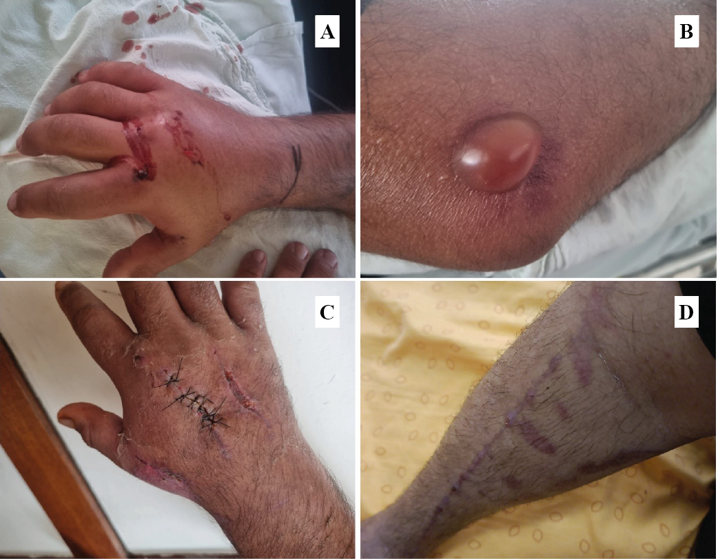
Figure 2. Figure 2. A) Edema and B) bullae along the right upper limb of a patient bitten by a Mesoamerican rattlesnake (Crotalus simus). Photographs were taken on the second day after the bite. C) Recovery of the hand bitten by a Mesoamerican rattlesnake (Crotalus simus) and D) fasciotomy scars on the right upper limb of the same patient. C) and D) Photographs were taken 14 days after envenoming and following the patient’s discharge.
On the sixth day, the patient underwent surgical washout and a partial wound closure. Additionally, the doctors placed a vacuum-assisted closure (VAC) therapy. The culture results were negative for aerobic microorganisms. However, on the seventh day, the infectious disease team escalated antibiotic therapy to ceftazidime and linezolid to prevent nosocomial infections, such as those caused by Staphylococcus aureus and Pseudomonas species. The same day, the patient also developed a pruritic erythematous maculopapular rash on the back, gluteal region, and groin, consistent with serum sickness secondary to antivenom administration. Dexamethasone 8 mg was administered STAT, followed by 4 mg every 12 h for 3 days.
On the tenth day, VAC therapy device was removed and a progressive improvement in laboratory parameters was observed (Table 1). On the 14th day, antibiotic therapy was discontinued after 7 days, and the patient was discharged with antihistamines due to the persistence of the rash (Figure 2). From day 14 to day 37, the patient attended follow-up appointments with the reconstructive surgery team, during which wound care was provided, sutures were removed, and a 1 cm² skin defect on the dorsum of the hand—located near the second metacarpophalangeal joint—was closely monitored. By this time, the wounds were almost completely healed, although residual edema and limited hand mobility persisted. After discharge, the patient also underwent several rehabilitation procedures, including physical and occupational therapy provided through the CCSS and 20 sessions of hyperbaric oxygen therapy at his own expense. These interventions resulted in significant functional improvement. However, the patient was left with physical sequelae, with approximately 90% recovery of mobility in the right upper limb. Additionally, altered sensitivity characterized by hyperalgesia and allodynia persists, which continues to be managed through specialized therapy.
Discussion
Throughout the clinical picture of the patient, a mainly hemorrhagic envenomation affecting the coagulation processes could be observed. Snake venom Zn2+ metalloproteinases (SVMPs) are a group of proteins abundant in adult Mesoamerican rattlesnakes (~72%).11,12 These molecules are known to cause hemorrhagic effects by attacking components of the basement membrane and extracellular matrix.13 Therefore, SVMPs are associated with hemorrhage disturbances and a consumption coagulopathy. Additionally, adult C. simus venom is composed of ~5% serine proteinases.12 These toxins act on components of the of the coagulation cascade, on the fibrinolytic and kallikrein–kinin systems and on cells, ultimately causing destabilization in the hemostatic system.14 Therefore, the disorders in the coagulation times reported here are also associated with the presence of these molecules.
Crotalus envenomation is a well-documented topic.15 Venom composition in the genus is divided into type I venoms, characterized by a high content of SVMPs that produce a hemorrhagic clinical picture, and type II venoms, primarily composed of crotoxin, which produce a neurotoxic clinical picture.11,16 In this case, adult C. simus envenomation resulted in altered hematological and coagulation values. Significant hematological abnormalities after envenomation has also been found in C. culminatus and C. molossus,17,18 while coagulopathy as one of the main manifestations has been previously described for C. atrox and juvenile C. durissus terrificus.19,20 Conversely, neurological manifestations have been reported in C. cerastes, C. scutulatus, and some subspecies of C. durissus.21-23
During hospitalization, surgical management was emphasized. The reconstructive surgery service at Hospital Calderón Guardia diagnosed the patient with compartment syndrome, which led to a fasciotomy and several surgical washouts. Although crotaline envenomation can manifest symptoms suggestive of compartment syndrome, clinical evidence indicates that true compartment syndrome is rare and should be confirmed through carefully assessed serial compartment pressure measurements.24,25 Therefore, the use of therapeutic interventions such as fasciotomy is discouraged because it increases the morbidity of snakebite envenomation and prolongs hospitalization.25-27 The persistence of edema should have been critically evaluated, and additional evidence, such as necrotizing bacterial fasciitis or myositis,28,29 should have been considered prior to performing the surgical intervention.
The neutralization of C. simus venom by PoliVal-ICP is expected, as this antivenom consists of purified horse immunoglobulins derived from a mixture of venoms from adult B. asper, Lachesis stenophrys, and C. simus.11,30 The early and timely approach with antivenom was fundamental for the reduction of tissue damage and major hemorrhagic complications, besides being the most important factor in saving the patient’s life and limb. Despite the persistence of edema, the doctors did not recommend administering an additional third dose of antivenom. Most snakebite cases in Costa Rica report satisfactory use of 10 (52%) and 15 (24%) ampoules;6 while in this case, the patient had already received 20 vials. The return of biochemical values to normal ranges supports the doctors’ decision not to provide additional antivenom.
The use of antibiotics in snakebite treatment is common to prevent secondary infections caused by bacteria present in the animal’s oral cavity.25 The use of adjunctive antibiotic therapy following a snakebite has been reported in all documented cases in Costa Rica, although without specific protocols.6 Immediate empirical treatment with a broad-spectrum antibiotic is justified if bite wounds are contaminated, warm, necrotic, or if erythema persists.25 Therefore, the initial use of cefotaxime and metronidazole owing to the delayed resolution of inflammation and sustained erythema was beneficial for the patient. Moreover, the performance of multiple surgical interventions led to an antibiotic escalation to ceftazidime and linezolid to prevent nosocomial infections, a standard protocol in hospitals across the country.
Finally, this case describes the event, evolution, and management of a 40-year-old patient weighing 100 kg with associated comorbidities: hypertension, dyslipidemia, and glaucoma. He was bitten by a male C. simus measuring 140.5 cm in length and weighing 1650 g. The patient was treated according to the established protocols in Costa Rica for snakebite management, which included the timely administration of the antivenom specific to Central and South American vipers, PoliVal-ICP. He subsequently required specialized medical care at a hospital and survived severe envenomation. This case demonstrates a successful outcome, despite the challenges faced by the Costa Rican social security system.
References
1. Gutiérrez JM, Calvete JJ, Habib AG, Harrison RA, Williams DJ, Warrell DA. Snakebite envenoming. Nat. Rev. Dis. Primers. 2017; 3: 17063. DOI: 10.1038/nrdp.2017.63
2. Harrison RA, Hargreaves A, Wagstaff SC, Faragher B, Lalloo DG. Snake envenoming: a disease of poverty. PLoS Negl. Trop. Dis. 2009; 3: e569. DOI: 10.1371/journal.pntd.0000569
3. Kasturiratne A, Wickremasinghe AR, De Silva N, Gunawardena NK, Pathmeswaran A, Premaratna R, et al. The global burden of snakebite: a literature analysis and modelling based on regional estimates of envenoming and deaths. PLoS Med. 2008; 5: e218. DOI: 10.1371/journal.pmed.0050218
4. Hansson E, Sasa M, Mattisson K, Robles A, Gutiérrez JM. Using geographical information systems to identify populations in need of improved accessibility to antivenom treatment for snakebite envenoming in Costa Rica. PLoS Negl. Trop. Dis. 2013; 7: e2009. DOI: 10.1371/journal.pntd.0002009
5. Sasa M, Vázquez S. Snakebite envenomation in Costa Rica: a revision of incidence in the decade 1990–2000. Toxicon. 2003;41:19–22. DOI: 10.1016/s0041-0101(02)00172-1
6. Sasa M, Segura-Cano SE. New insights into snakebite epidemiology in Costa Rica: A retrospective evaluation of medical records. Toxicon X. 2020; 7: 100055. DOI: 10.1016/j.toxcx.2020.100055
7. Chippaux JP. Incidence and mortality due to snakebite in the Americas. PLoS Negl. Trop. Dis. 2017; 11: e0005662. DOI: 10.1371/journal.pntd.0005662
8. Fernández P, Gutiérrez JM. Mortality due to snakebite envenomation in Costa Rica (1993–2006). Toxicon. 2008; 52: 530-533. DOI: 10.1016/j.toxicon.2008.06.018
9. Gutiérrez JM. Snakebite poisoning in Latin America and the Caribbean: an integral view from a regional perspective. Bol. Mal. Salud Amb. 2011; 51: 1-16.
10. Bolaños R. Las serpientes venenosas de Centroamérica y el problema del ofidismo. Rev. Costarric. Cienc. Méd. 1982; 3: 165-184.
11. Calvete JJ, Sanz L, Cid P, De La Torre P, Flores-Díaz M, Dos Santos MC, et al. Snake venomics of the Central American rattlesnake Crotalus simus and the South American Crotalus durissus complex points to neurotoxicity as an adaptive paedomorphic trend along Crotalus dispersal in South America. J. Proteome Res. 2010; 9: 528–544. DOI: 10.1021/pr9008749
12. Lomonte B, Fernández J, Sanz L, Angulo Y, Sasa M, Gutiérrez JM, et al. Venomous snakes of Costa Rica: biological and medical implications of their venom proteomic profiles analyzed through the strategy of snake venomics. J. Proteomics. 2014; 105: 323–339. DOI: 10.1016/j.jprot.2014.02.020
13. Escalante T, Rucavado A, Fox JW, Gutiérrez JM. Key events in microvascular damage induced by snake venom hemorrhagic metalloproteinases. J. Proteomics. 2011; 74: 1781–1794. DOI: 10.1016/j.jprot.2011.03.026
14. Serrano SMT. The long road of research on snake venom serine proteinases. Toxicon. 2013; 62: 19–26. DOI: 10.1016/j.toxicon.2012.09.003
15. Boyer LV, Seifert SA, Clark RF, McNally JT, Williams SR, Nordt SP, et al. Recurrent and persistent coagulopathy following pit viper envenomation. Arch. Intern. Med. 1999; 159: 706-710. DOI: 10.1001/archinte.159.7.706
16. White J. Snake venoms and coagulopathy. Toxicon. 2005; 45: 951-967. DOI: 10.1016/j.toxicon.2005.02.030
17. Hardy DL, Jeter M, Corrigan, JJ. Envenomation by the northern blacktail rattlesnake (Crotalus molossus molossus): report of two cases and the vitro effects of the venom on fibrinolysis and platelet aggregation. Toxicon. 1982; 20: 487-493. DOI: 10.1016/0041-0101(82)90012-5
18. Zarzosa V, Lomonte B, Zamudio F, Ponce-López R, Olvera-Rodríguez F, Borja M, et al. Venom of the neotropical rattlesnake, Crotalus culminatus: Intraspecific variation, neutralization by antivenoms, and immunogenicity in rabbits. Biochimie. 2024; 216: 160-174. DOI: 10.1016/j.biochi.2023.10.014
19. Bucaretchi F, De Capitani EM, Branco MM, Fernandes LCR, Hyslop S. Coagulopathy as the main systemic manifestation after envenoming by a juvenile South American rattlesnake (Crotalus durissus terrificus): Case report. Clin. Toxicol. 2013; 51: 505–508. DOI: 10.3109/15563650.2013.802796
20. Budzynski AZ, Pandya BV, Rubin RN, Brizuela BS, Soszka T, Stewart GJ. Fibrinogenolytic afibrinogenemia after envenomation by western diamondback rattlesnake (Crotalus atrox). Blood. 1984; 63: 1-14. DOI: 10.1182/blood.V63.1.1.1
21. Bosak AR, Ruha AM, Graeme KA. A case of neurotoxicity following envenomation by the Sidewinder rattlesnake, Crotalus cerastes. J. Med. Toxicol. 2014; 10: 229-231. DOI: 10.1007/s13181-013-0373-0
22. Frare BT, Silva-Resende YK, Dornelas BC, Jorge MT, Souza-Ricarte VA, Alves LM, et al. Clinical, laboratory, and therapeutic aspects of Crotalus durissus (South American Rattlesnake) victims: A literature review. Biomed Res Int. 2019; 2019: 1345923. DOI: 10.1155/2019/1345923
23. Smelski G, Cardwell M, Larsen J. Neurotoxic respiratory failure absent following Arizona rattlesnake bites. Toxicon. 2023; 224: 107034. DOI: 10.1016/j.toxicon.2023.107034
24. Cañas CA. Is the acute compartment syndrome diagnosed in snake bites true?: A review. Medicine. 2024; 103: e40008. DOI: 10.1097/md.0000000000040008
25. Warrell DA, Williams DJ. Clinical aspects of snakebite envenoming and its treatment in low-resource settings. Lancet. 2023; 401: 1382-1398. DOI: 10.1016/S0140-6736(23)00002-8
26. Cumpston KL. Is there a role for fasciotomy in Crotalinae envenomations in North America?. Clin. Toxicol. 2011; 49: 351-365. DOI: 10.3109/15563650.2011.597032
27. Darracq MA, Cantrell FL, Klauk B, Thornton SL. A chance to cut is not always a chance to cure-fasciotomy in the treatment of rattlesnake envenomation: a retrospective poison center study. Toxicon. 2015; 101: 23-26. DOI: 10.1016/j.toxicon.2015.04.014
28. Banda CH, N’gambi C. Necrotizing fasciitis with mediastinitis following snakebite envenomation. SAGE Open Med. Case Rep. 2019; 7: 2050313X18823090. DOI: 10.1177/2050313X18823090
29. Yuenyongviwat V, Laohawiriyakamol T, Suwanno P, Kanjanapradit K, Tanutit P. Calcific myonecrosis following snake bite: a case report and review of the literature. J. Med. Case Rep. 2014; 8: 193. DOI: 10.1186/1752-1947-8-193
30. Alfaro-Chinchilla A, Segura A, Gómez A, Díaz C, Corrales G, Chacón D, et al. Expanding the neutralization scope of the Central American antivenom (PoliVal-ICP) to include the venom of Crotalus durissus pifanorum. J. Proteomics. 2021; 246: 104315. DOI: 10.1016/j.jprot.2021.10431

Esta obra está bajo una licencia internacional: Creative Commons Atribución-NoComercial-CompartirIgual 4.0.